

Let’s dive into the different layers of the eye and see what gives us this amazing thing we call sight. I’m going to nerd this one out and take you right back to the classroom. The organ of vision is the most important of all the human senses because a person receives about 80% of information about the outside world through the eyes. It really is a truly amazing sense if you’re lucky enough to have it.
Eyeball
The eye is located in the orbit and is surrounded by soft tissues (adipose tissue, muscles, nerves, etc.). It is covered in front with conjunctiva. The eyeball consists of three layers, limiting the inner space to the anterior, posterior chamber of the eye, as well as the space filled with the vitreous body – the vitreous chamber.
The outer layer of the eye
It is represented by dense connective tissue. It consists of a transparent cornea in the anterior part of the eye and a white opaque sclera over the rest of the length. Possessing elastic properties, these two shells form the characteristic shape of the eye.
Cornea
This is the transparent part of the outer layer of the eye. The place of its transition to the sclera is called the limbus. The shape of the cornea is ellipsoidal, vertical diameter – 11 mm, horizontal – 12 mm. The thickness of the cornea is about 1mm. The transparency of the cornea is explained by the uniqueness of its structure, in it, all cells are located in a strict optical order and there are no blood vessels in it.
The cornea consists of 5 layers:
1. The front epithelium
2. The bowmen’s membrane;
3. Stroma
4. Descemet’s shell
5. The back epithelium (endothelium)
The cornea is rich in nerve endings; therefore, it is very sensitive. The cornea not only transmits but also refracts light rays, it has great refractive power.
Sclera
This is the opaque part of the outer layer of the eye, which is white in color. Despite its thickness of 1 mm, it is very dense in nature. The sclera consists mainly of dense fibers, which give its strength. The muscles of the eye are attached to the sclera.
Middle layer or vascular layer
This is the middle layer of the eye, consisting mainly of blood vessels of different calibers.
Iris
It resembles a circle with a hole. The iris consists of 2 muscles: narrowing and widening the pupil with contraction and relaxation of which the size of the pupil changes. The composition of the iris includes cells containing pigment, which determine the color of the eyes. The iris performs the function of regulating the width of the light beam that falls inside the eye.
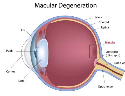
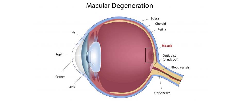
Inner layer or Retina
In the retina, light is converted into nerve impulses that are transmitted through the nerve fibers to the brain. There they are analyzed, and the person perceives the image. The retina consists of 6 layers. The outer layer of the retina is pigmented. It absorbs light, reducing its dispersion inside the eye. In the next layer are the processes of cells of the retina – rods, and cones. The processes contain visual pigments – rhodopsin (rods) and iodopsin (cones).
Thank you so much for taking the time and getting yourself better acquainted with this miracle we call the eye. With so much that the eye does it’s no wonder this little organ is so complex. Another reason why I have spent my entire academic life dedicated to learning about how to keep your eyes healthy and vision correct. Please visit an eye doctor today and keep this little miracle of yours healthy for years to come.


